Neuroscience 101

It is in your head

Monthly Archives: November 2010

Haiti’s Cholera Outbreak

This Holiday, add them to your list! They need us.

Donate –  Save The Children – http://tinyurl.com/3acl5gw

Hyperlipidemia

What is hyperlipidemia?

  • Lipoprotein lipase is located in the endothelium of adipose and muscle capillaries, this enzyme is responsible for the release of free fatty acids from both VLDLs and chylomicrons. It is inhibited by Apo CIII and activated by Apo CII. Lipoprotein lipase is responsible for the breakdown of triglycerides to free fatty acids. The process does not require insulin, unlike the uptake of free fatty acids into muscle or adipose tissue. MTP is involved in Apo B assembly. LCAT esterifies free cholesterol into hydrophobic cholesterol esters that move into the center of HDL particles. Cubulin is the Apo AI receptor responsible for renal clearance of HDL.
  • Although most hepatic receptors for triglyceride-rich lipoproteins will recognize Apo E, allowing clearance though the liver, Apo B100 is required for LDL binding to its receptor in the liver. Mutations within the gene for this lipoprotein can cause a form of familial hypercholesterolemia. Most VLDL and IDL also contain Apo E, as well as Apo B100. It is only LDL that is dependent on Apo B100 for hepatic uptake secondary to its lack of Apo E.
  • Epidemiological and clinical studies provide evidence that HDL-C levels are linked to rates of coronary events. The cardioprotective effects of HDL-C have been attributed to its role in reverse cholesterol transport. The cardioprotective effects of HDL-C have been attributed to its effect on endothelial cells and its antioxidant activity. Low HDL-C is caused by cigarette smoking, very high carbohydrate intake, and the use of certain drugs (e.g., progestational agents, anabolic steroids). Low HDL-C is one of five major CHD risk factors. HDL-C level is a component of the Framingham scoring system, the method used to estimate 10-year CHD risk and to determine the intensity of lipid lowering therapy. While the new guidelines do emphasize the importance of HDL in the management of hyperlipidemia, they have not set specific targets at this time because we do not yet have appropriate data from a large randomized controlled trial.

Case I: A 35-year-old African American male presents to your office for follow up on his free cholesterol screening that he received at the mall this past weekend. He is a nonsmoker who takes no medication and reports that he has been relatively healthy for his entire life except for occasional flares of gout. He has a grandfather who had an MI at 60. He exercises with a trainer four times per week and appears to be in good physical shape.

Total Cholesterol 151
Triglycerides 130
HDL-C 25
LDL-C 100

Based on current studies, which therapy would most benefit your patient at this time?

Answer: With a statin you can expect to raise the HDL-C by 5-15%.

Based on the current guidelines, this patient’s LDL is at/below goal. The most obvious abnormality in his profile is the markedly low HDL. Initiating therapeutic lifestyle changes that incorporate weight loss and increased physical activity would be the first step taken to raise HDL; however, this particular patient is already exercising on a regular basis. His goal HDL is 50, which may be unobtainable. Normally, trying to raise HDL via drug therapy, niacin and statins are usually excellent choices. In this particular patient, however, niacin would not be the best choice, as it tends to exacerbate gout.

Case II: A 31-year-old man seeks medical advice after his older brother survives a myocardial infarction. He has no medical history, does not smoke or drink alcohol. His BP is 130/85. He has corneal arcus and an Achilles tendon xanthoma. His lipid profile shows total cholesterol of 300 mg/dL with an LDL-C of 240 mg/dL and an HDL of 40 mg/dL. Which phenotypical lipid disorder is this patient expressing?

Answer: Type II hyperlipidemia or familial hypercholesterolemia is due to defects in LDL uptake into the liver secondary to problems with the LDL receptor. Heterozygotes have a prevalence of 1:500 and manifest coronary disease by the 4th decade. Xanthomas, a positive family history, and elevated LDL make the diagnosis.

Case III: A 45-year-old man is seeking treatment for his elevated cholesterol found at a free health fair. He is asymptomatic and has no physical signs of hypercholesterolemia such as corneal arcus or xanthomas. His fasting cholesterol profile shows a total cholesterol of 220 mg/dL, triglycerides 400 mg/dL, LDL 100 mg/dL and HDL 40 mg/dL. A postprandial triglyceride level is 1000 mg/dL. You suspect that he has familial hypertriglyceridemia (Type IV hyperlipoproteinemia). The metabolic defect responsible for his condition is?

AnswerIncreased hepatic production of VLDL. Decreased LDL receptors cause Type II hyperlipidemia or familial hypercholesterolemia. Abnormal Apo B100 causes a phenotype indistinguishable from familial hypercholesterolemia. Decreased LPL activity causes familial hyperchylomicronemia. Abnormal Apo E is the metabolic defect in Type III hyperlipoproteinemia.

  • Although familial combined hyperlipidemia has been defined in the laboratory as a total cholesterol greater than the 90th percentile and/or a plasma triglyceride level greater than the 90th percentile, both adjusted for age and gender, these cutpoints could miss up to 25% of affected patients. Apolipoprotein B markers can be added to total cholesterol or triglyceride levels to better predict familial combined hyperlipidemia. Apo B levels greater than 1200 mg/dL greatly increase the likelihood of familial combined hyperlipidemia in patients with elevated triglycerides (>1.5 mmol/L) or total cholesterol (>6 mmol/L).

Case IV: A 42-year-old female consults you for advise on staying healthy. She exercises daily and has never had any symptoms of heart disease. She has never smoked. Her father died of a massive myocardial infarction at the age of 44. She has been treated for hypertension over the last five years with an angiotensin converting enzyme inhibitor (ACE) and says that her BP is usually 140/88. Her total serum cholesterol is 220 mg/dl and her HDL-C is 30.

You order a complete fasting lipid profile and the results come back as follows:

Total Cholesterol 230 mg/dl
Triglycerides 455 mg/dl
HDL-C 34 mg/dl

Calculate the patient’s LDL-C?

Answer: You are unable to calculate it given the current information because the formula for the calculation of LDL-C:LDL-C = Total Cholesterol – HDL-C – (Triglycerides/5)

P.S. ATP III guidelines suggest for individuals who have 2+ risk factors that their LDL-C goal is <130 mg/dl. Her LDL-C goal is supposed to be <130 mg/dl.

Let’s just say her LDL-C is 165 mg/dl. Can we recommend her  a treatment based on the data provided? Nope because the ATP III guidelines state that the treatment to achieve the LDL-C goals in patients with 2+ risk factors should not only be based on the LDL-C levels but should also be further modified by the Framingham 10-year CHD Risk Calculation for these individuals.

Treatment Guide:

I. Raises HDL-C, lowers both LDL-C and triglycerides. No large randomized control trials have been reported for its use in isolated low HDL-C patients Niacin
II- Can raise HDL-C by 10-20%, modestly lower LDL-C and substantially lower triglycerides. Absolutely contraindicated in patients with severe renal or hepatic disease Fibrates
III- Apo-I Milano administreted weekly to 123 patients via IV infusion post ACS demonstrated a 1.06% decrease in atheroma volume as evaluated by intravascular ultrasound at five weeks. Exogenous HDL Mimetic
IV- Recommended by ATP III and the treatment of choice by raising HDL especially in patients with the metabolic syndrome. Increased physical activity.
V- Effectively lower LDL-C while mildly increasing HDL and decreasing triglycerides minimally. They are contraindicated in patients with active liver disease. Statins

Case V:

A 34-year-old obese male with a history of hypertension presents to your office for his first visit. He has a strong family history of CAD and currently has cut back to smoking two cigarettes a day. He is currently on no lipid lowering medications:

Total Cholesterol 200 mg/dl
Triglycerides 205 mg/dl
HDL-C 40 mg/dl
LDL-C 119 mg/dl

Answer: You initiate TLC(Therapeutic Lifestyle Changes), together with close follow up, are the initial steps in the treatment of moderate risk patients.

The patients returns for follow/up four months later and states that he has been watching his diet and that he now exercises three times per week. His laboratory work is listed below:

Total Cholesterol 195 mg/dl
Triglycerides 200 mg/dl
HDL-C 42 mg/dl
LDL-C 113 mg/dl

See, this time TLC didn’t help. We need to treat him with low dose statin. Based on the updated ATP III guidelines, this patient is considered to be in the moderate risk category (2+ risk factors and a 10-year risk of 10-20%). The new guidelines state that his LDL-C should be below 130 mg/dl and they give you the therapeutic option to lower it to treat the LDL-C to <100 mg/dl when the initial untreated measurements are between 100 and 129 mg/dl.

Pro tip: -hs-CRP- (high-sensitivity CRP): Elevated hs-CRP levels have been demonstrated to be effective in predicting the development of hypertension and a good predictor of incident type 2 diabetes and add prognostic values on vascular risk at all levels of the metabolic syndrome. hs-CRP levels can effectively be lowered with “statin” therapy. hs-CRP continues to evolve as a major risk de facto marker for CHD. In addition, it has emerged as a predictor of disease progression in many of the comorbidities associated with CHD: hypertension, type 2 diabetes mellitus. It has been demonstrated that hs-CRP can be reduced with statins and other antinflammatory agents.

Hyperlipidemia and Risks

  • CD40 ligand is a proinflammatory mediator that plays a crucial role in the pathogenesis of the acute coronary syndrome and has been shown to be elevated in patients with unstable coronary syndromes. In the MIRACL patient population, soluble CD40 ligand (sCD40L) was measured at baseline and at 16 weeks. Patients with an elevated sCD40L >90th centile compared to patients with a sCD40L <90th centile had an odds ratio of 1.86 for a recurrent event. Treatment with atorvastatin abolished this increased risk, it appears that only a high level of sCD40L is associated with increased cardiovascular risk. Even in the >90th centile patient group, event rates with atorvastatin treatment were similar to the <90th centile group. In addition, in the patient group with sCD40L <90th centile, treatment with atorvastatin was not associated with any decrease in event rate. This may allow selection of high-risk patients in whom early intensive statin therapy would decrease event rates.
  • Peroxisome proliferator-activated receptors (PPARs) are members of the nuclear receptor family that act as transcription factors involved in the regulation of several metabolic pathways that may influence cardiovascular risk. PPARs have three types. PPAR-alpha was the first discovered and controls target genes such as HMG-CoA-synthetase, Apo A1 and Apo-CIII. It is also the target for the fibric acid derivatives, gemfibrozil and fenofibrate. It was shown in PPAR-alpha deficient mice that the agonist of PPAR-alpha did not lower triglyceride levels. PPAR-gamma regulates the expression of acyl-CoA-synthetase, lipoprotein lipase and fatty acid binding protein. It is the target for the thiazolidione class of antidiabetic drugs. Its over-expression can drive the differentiation of fibroblasts to an adipocyte-like phenotype. Another activator of PPAR-gamma is oxidized linoleic acid, a component of oxidized LDL. This is one potential mechanism for the deleterious effects of oxidized LDL on plaque stability. With the changing of a fibroblast to an adipocyte-like phenotype, the “fibrous” cap is more susceptible to rupture.

Those information provided was for my healthcare colleagues and now, dumbing it down for the rest of you:

Wud, wud, wud? Hyperlipidemia is an elevation of lipids in the bloodstream, cholesterol, cholesterol esters, estersphospholipids and triglycerides.

Causes? Primary and secondary, primary causes: heredity and taking certain medications, however the greatest risk factor is a poor diet, with a fat intake >49% of total calories, saturated fat intake >10% of total calories and cholesterol intake >300 mg/day. Secondary causes may be a clue to the presence of an underlying systemic disorder. A convenient way to recognize secondary causes is to think of the four Ds(diet, drugs, disorders of metabolism, diseases). For example, hypothyroidism is the most common secondary cause of hyperlipidemia, also pregnancy, alcohol, estrogens, steroids.

Symptoms? No symptoms. Regular cholesterol screening with blood tests should be part of your life.

Treatment? As I mentioned above, TLC – Therapeutic Lifestyle Changes. Dietary and lifestyle modification, followed by drug therapy(which I gave details above above), if necessary.

Preventation: Exercise 3 times/week and watch your fat intake.

Have a great week. Happy early Thanksgiving!

Would you mind checking out this website and donate to the Haiti Cholera Children in Emergency Fund to support Save the Children’s response to the deadly cholera outbreak in Haiti? Thanks in advance.

https://secure.savethechildren.org/site/c.8rKLIXMGIpI4E/b.6238941/k.E6AE/Support_the_Haiti_Cholera_Children_in_Emergency_Fund/apps/ka/sd/donor.asp?c=8rKLIXMGIpI4E&b=6238941&en=cvINK3PLLbLWJdNOIaLSIfM5IvL5KcPWKmKZKgMZLnJUIfPaG

Medically Relevant: Something

"The sound of the waves collide..." - Deftones

"If we're still alive, my regrets are few. If my life is mine, what shouldn't I do?" - Metric

"I see her everywhere, long amber hair, calling out my name" - The Black Keys

"And in my darkest moment, fetal and weeping, the moon tells me a secret, her confidant "as full and bright as I am, this light is not my own and a million light reflections pass over me. The source is bright and endless, she(Amber) resuscitates the hopeless, without her we are lifeless satellites drifting."" - Tool

"Any way the wind blows" - Queen

Case Study: Active Rheumatoid arthritis

A 28-year-old woman has noted pain and swelling of her hands and wrists for six months. She has discomfort with walking, notes fatigue and difficulty using the keyboard at work and is having difficulty caring for her 2-year-old baby. She has been taking diclofenac 150 mg/day. Her exam shows tenderness and swelling of her PIP, MCP joints and wrist joints, as well as knee swelling. The MTP joints are also tender on squeeze. The labs include a negative rheumatoid factor, sed rate of 45 mm/hour and Hct of 36.


Radiograph.


Which of the following would you do now?

  • Repeat rheumatoid factor.
  • Order MRI of hands.
  • Start prednisone 30 mg/day.
  • Talk to her about starting methotrexate therapy.
  • Refer to occupational therapy.

Answer: Talk to her about starting methotrexate therapy and refer to occupational therapy.

Explanation: This patient has active RA and needs to be started on a DMARD therapy. A referral to occupational therapy for a hand evaluaton, including hand exercises and adaptive equipment and splints, is also indicated to help improve her activity level.

The decision about starting a DMARD is not dependent on laboratory findings such as the rheumatoid factor status, acute phase response or the radiographic appearance. You should not wait for the presence of erosions as the indication to start a DMARD. The decision about starting a DMARD should be based on the clinical activity of the disease. Which DMARD you recommend should take into account disease activity, functional status, life style issues and the underlying health status of the patient. For this young woman, the DMARD methotrexate is appropriate based on the disease activity and functional disability.

There is no indication to order an MRI to look for erosions since the decision to start a DMARD should be made based on clinical activity. Similarly, it does not matter if you do a repeat rheumatoid factor because it will not impact on the decision to start a DMARD. Steroids might be added to the regimen, but the dose is too high to use as a bridge while you wait for the methotrexate to work.

Neuroscience: A few Major Depression Disorder facts

  • About 60% of patients with MDD are female.
  • Patients have a markedly diminished interest or pleasure in all, or almost all, activities (anhedonia), one of the cardinal symptoms of MDD.
  • About 30-35% of patients with MDD will develop a sufficient number of recurrences over time such that the illness is considered chronic.
  • Grossly disorganized or catatonic behavior is a characteristic symptom of schizophrenia and is not a diagnostic criterion for MDD.
  • Fatigue or loss of energy is also a diagnostic criterion for MDD.
  • Sexual and physical abuse in childhood, as well as parental neglect, are risk factors for MDD.
  • In about 90% of suicides, a psychiatric diagnosis is possible with about 60% being MDD.
  • Male suicides outnumber female suicides by about 4:1.
  • Annually, there are about 50% more suicides than homicides.
  • Suicide is the 11th leading cause of death in the United States.
  • Various indices of HPA activity, such as the dexamethasone suppression test, have been used to demonstrate HPA overactivity in a substantial percentage of patients with MDD.
  • Although Parkinson’s Disease and Alzheimer’s Disease are risk factors for depression, it is unlikely that their association with MDD is stress-related.
  • Loss of parents at an early age is an established risk factor for MDD.
  • Increased adrenal and pituitary size have been found in several studies, possibly due to chronic hyperactivity of the HPA axis.
  • Glucocorticoid antagonists have not yet been found to be effective clinically, although there is great interest in them based on preclinical data.
  • There is a high degree of alcohol and drug abuse comorbidity and this may be stress-related.
  • Reduced hippocampal volume is likely due to deleterious effects of elevated glucocorticoids in MDD on the hippocampus.
  • Chest pain and discomfort are symptoms of panic disorder and are not used to make the diagnosis of MDD.
  • Avoidance of situations with unfamiliar people is a symptom of social anxiety disorder and not one used to make the diagnosis of MDD.
  • The substantia nigra gives rise to dopaminergic, not noradrenergic, neurons and is not known to be altered in depression.
  • Low levels of the serotonin metabolite, 5-hydroxyindoleacetic acid (5-HIAA) in CSF was an early finding that was confirmed and stimulated the idea that 5-HT was involved in depression.
  • After it was discovered that TCA (imipramine) was found to block NE reuptake, it was soon noted that it also blocked the uptake of 5-HT, which contributed to the view that antidepressants worked by affecting serotonergic transmission.
  • Reserpine depletes the brain of 5-HT and NE. When coupled with the idea that the drug produced depression in certain hypertensive patients, this fact also contributed to the idea of a serotonin or norepinephrine deficiency in depression.
  • Serotonergic cell bodies in the raphe nuclei in the midbrain send serotonergic projections to these areas. Neither sertraline nor nefazodone enhance noradrenergic transmission. Since fluoxetine acts through 5-HT (i.e., it is an SSRI), this did not occur.
  • Some patients treated with reserpine for hypertension developed depression and this influenced the view that monoamine deficiency contributed to depression.
  • Subsequent reanalysis of the data indicated that this occurred primarily in susceptible individuals, particularly those with a prior history of depression.
  • Stress activates noradrenergic cell bodies in the locus coeruleus, another contributiion to the view that the central noradrenergic system is involved in processes relating to activation, alerting and vigilance.
  • Postmortem studies demonstrated that abnormal amounts of noradrenergic proteins, such as the NE synthetic enzyme, tyrosine hydroxylase, are found in the brains of patients with major depression.
  • The low 5-HIAA was subsequently found to be more related to the risk for suicide rather than MDD per se.
  • Fluoxetine is an SSRI and 5-HT is necessary for its beneficial clinical effects.
  • The striatum receives little, if any, noradrenergic innervation.
  • Low levels of MHPG were found.
  • GABA is the primary inhibitory transmitter in brain.
  • HT is not metabolized by COMT, although catecholamines are.
  • Early studies found that tryptophan and 5-hydroxytryptophan, which are precursors of 5-HT, may have mild antidepressant-like activity, but this remains controversial. The early results, though, further substantiated a role for 5-HT in the treatment of MDD.
  • Noradrenergic cell bodies in the midbrain, particularly the locus coeruleus, send projections to these areas.
  • Serotonergic cell bodies in the raphe nuclei in the midbrain also send serotonergic projections to these areas. SSRIs are drugs that selectively block the reuptake of 5-HT and are the most widely prescribed drugs for MDD.
  • Low levels of the serotonin metabolite, 5-HIAA, were associated with an increased risk of violent suicide.
  • Imipramine blocks the uptake of NE as well as 5-HT, whereas fluoxetine selectively blocks the uptake of 5-HT.
  • The reduced hippocampal volume is likely due to deleterious effects of elevated glucocorticoids in MDD on the hippocampus.
  • Although it is true that monoamine oxidase inhibitors (MAOIs) such as phenelzine raise the concentration of dopamine in brain, this is not relevant for an involvement of NE or 5-HT in antidepressant action. There is no evidence to support any aspect of the statement that serotonin activates central neurons containing substance P so as to reduce emotional “pain” in depressives.
  • Amphetamine has mood-elevating effects attributed to its ability to facilitate the release of catecholamines, NE and dopamine.
  • Tryptophan hydroxylase is the rate-limiting enzyme in 5-HT synthesis.
  • Desipramine and nortriptyline are selective inhibitors of NE reuptake.
  • Early studies that found low levels of the NE metabolite, 3-methoxy-4-hydroxyphenylglycol (MHPG), in the urine of depressed patients contributed to the NE deficiency hypothesis of depression.
  • Various types of antidepressants (TCAs, MAOIs, SSRIs) all enhance transmission caused by either NE or 5-HT and is a primary reason for thinking that these transmitters are involved in antidepressant action.
  • The primary mechanism for terminating the activity of NE or 5-HT in the synapse is not MAO but rather the transporters for these biogenic amines.
  • BBH is involved in thesynthesisof NE.
  • Desipramine exerts its therapeutic effect by enhancing noradrenergic transmission. In depressives, an increased risk of violent suicide is not associated with low levels of the dopamine metabolite, homovanillic acid (HVA) in CSF. Rather, as I mentioned previously, low levels of the serotonin metabolite, 5-HIAA, were associated with an increased risk of violent suicide.
  • Reserpine lowers brain concentrations of NE and 5-HT. Even though it is not an uptake inhibitor, mirtazapine has antidepressant activity linked to its ability to enhance noradrenergic transmission by blocking inhibitory noradrenergic autoreceptors, i.e., it is a potent α2 adrenergic receptor antagonist and α2 adrenoceptors are adrenergic autoreceptors.
  • The mechanism underlying bupropion’s efficacy in depression is not well understood but is unlikely to involve 5-HT.
  • Nortriptyline is a selective NE reuptake inhibitor.
  • By inhibiting the catabolism of NE by MAO in the neuron, phenelzine enhances noradrenergic transmission.
  • Fluoxetine is an SSRI and does not directly affect noradrenergic transmission.
  • Lithium is thought to act by affecting signal transduction processes and not noradrenergic transmission.
  • By blocking the re-uptake of NE, imipramine enhances noradrenergic transmission.
  • Nefazodone‘s mechanism of action is unclear. It is a very weak inhibitor of NE reuptake in vitro and is unlikely to produce this effect clinically.
  • Sertraline is an SSRI and does not directly affect noradrenergic transmission.
  • There is some debate as to whether venlafaxine inhibits the reuptake of NE at all doses used clinically or only at higher doses (e.g., >150-225 mg daily). Certainly, at higher doses, it does block the reuptake of NE and would enhance noradrenergic transmission.
  • Clozapine does not block the reuptake of NE but rather has α1 adrenoceptor antagonist activity.
  • Chlordiazepoxide does not enhance noradrenergic transmission as it does transmission mediated by GABA. As a selective and potent inhibitor of NE reuptake, desipramine enhances noradrenergic transmission.
  • The preponderance of evidence indicates that at daily doses of <150 mg daily venlafaxine acts primarily as an SSRI. At higher doses, it blocks the reuptake of NE as well, so that it functions as a dual 5-HT/NE reuptake inhibitor.
  • The selectivity of escitalopram for the serotonin transporter is so high that it functions as an SSRI irrespective of dose.
  • The selectivity of maprotiline for the norepinephrine transporter is so high that it functions as a NE reuptake inhibitor irrespective of dose.
  • Imipramine acts as a dual inhibitor of 5-HT and NE reuptake irrespective of dose.
  • Tranylcypromine is a non-selective inhibitor of MAO-A and MAO-B and, as such, inhibits both irrespective of dose.
  • Selegiline inhibits MAO-B more potently than MAO-A. At lower doses, then, selegiline selectively inhibits MAO-B, while at higher doses it inhibits both MAO-A and MAO-B.
  • Nefazodone weakly inhibits both NE and 5-HT uptake and there are no data to indicate that its pharmacological effects are dose dependent.
  • Phenelzine is a non-selective inhibitor of both MAO-A and MAO-B and, as such, inhibits both irrespective of dose.
  • The selectivity of desipramine for the NE transporter is so high that it functions as a NE reuptake inhibitor irrespective of dose.
  • The selectivity of paroxetine for the 5-HT transporter is so high that it functions as an SSRI irrespective of dose.
  • Escitalopram does not block the uptake of dopamine. Slowing of the firing rate of serotonergic cell bodies does occur very rapidly after administration of an SSRI, presumably due to the increased 5-HT in the synapse acting on inhibitory autoreceptors (5-HT1A) on serotonergic cell bodies/dendrites so as to slow cell firing.
  • Increase in the concentration of serotonin in the extracellular space in the hippocampus would occur after several weeks of administration of an SSRI due to the combination of blockade of 5-HT reuptake and desensitization of 5-HT autoreceptors producing a “normal” firing rate of serotonergic neurons.
  • An SSRI would not be expected to influence the release of dopamine directly. Downregulation of βadrenergic receptors is produced over time by drugs that block the uptake of NE but not by SSRIs.
  • Blockade of the uptake of serotonin (5-HT) is the key initial pharmacologic effect caused by SSRIs.
  • The enhancement of serotonergic transmission caused by SSRIs would activate whatever 5-HT receptor would be present in the particular synapse, including 5-HT3 receptors.
  • Recent data suggest that a time dependent effect of all classes of antidepressants is an increase in neurogenesis in the dentate gyrus of the hippocampus.
  • Noradrenergic uptake inhibitors decrease the firing rate of noradrenergic cell bodies but SSRIs do not have any effect.
  • Desensitization of inhibitory 5-HT1A autoreceptors is an effect that permits the firing rate of serotonergic cell bodies to return towards normal over time following the initial suppression of firing rate caused by SSRIs.
  • There are no conclusive data indicating that one class of antidepressant acts more quickly than any other class. In terms of the likelihood of patients improving, ECT produces improvement in a greater percentage of patients than drug therapy. A recent meta-analysis indicates that this is true — 57% of the total drug/placebo difference in severity measured over six weeks of treatment occurred in the first two weeks.
  • In terms of overall efficacy in non-selected patients with MDD, different classes of antidepressant drugs all have comparable efficacy. Acute effects of the drugs on noradrenergic or serotonergic neurons trigger more slowly-developing effects that are thought to be involved in efficacy. Those drugs that block muscarinic cholinergic receptors, primarily TCAs, produce this effect very quickly. Stimulation of 5-HT3 serotonergic receptors occurs quickly as a consequence of inhibition of 5-HT reuptake and is thought to account for the nausea observed initially with SSRIs.
  • Different types of antidepressants produce this effect after 2-3 weeks of administration. Increase of brain neurotrophic factor (BDNF) is another effect that different types of antidepressants produce in 2-3 weeks. The increase in BDNF is thought to be involved in the increase in neurogenesis. Another effect caused by different types of antidepressants after 2-3 weeks of administration is the increase in CREB which may be involved in the increase in BDNF.
  • Antidepressants do not have pharmacological effects that enhance dopaminergic transmission in the striatum.
  • Antidepressants do not slow the firing rate of dopaminergic cell bodies but different types of antidepressants produce this effect on noradrenergic or serotonergic cell bodies.
  • Antidepressants do not increase the release of dopamine. In general, antidepressants have little direct effect on dopaminergic neurons.
  • Antidepressants that enhance noradrenergic transmission produce a time-dependent downregulation of β adrenergic receptors.
  • An increase in serotonergic neurotransmission would produce activation of all types of receptors for 5-HT, including the 5-HT1A receptor.
  • Antidepressants have little direct effect on dopaminergic neurons.
  • There are no data that indicate conclusively that one drug class of antidepressant differs from any other type in the onset of therapeutic improvement.
  • Drugs that do block H1 histamine receptors, TCAs and mirtazapine, do so very quickly after their administration. As mentioned previously, chronic treatment with SSRIs produces a desensitization of inhibitory 5-HT1A autoreceptors, not supersensitivity.

Taking a break: What have you learned so far? : Case

Ms. Smith, a 35-year-old woman, has been referred to you for psychiatric treatment. During the past month she appeared in the emergency room twice, each time complaining of chest pain, shortness of breath and dizziness. After an extensive medical examination, she was diagnosed as suffering from an “anxiety attack.” Ms. Smith describes to you her concern about dying, after the sudden death of her father from an MI six weeks ago. Other recent events in her life include the loss of her job and sadness after the break-up of a recent romance with a man living next door to her. She has been sleeping 12-16 hours per day for the last few months and has gained 35 pounds. She describes non-suicidal thoughts about death, loss of interest in activities, dysphoria and a low level of energy. Over the last month, she has experienced periods of intense anxiety which last for about 15 minutes, including the two which brought her to the emergency room. She describes herself as a recovering alcoholic, sober for 10 years. The patient has taken five doses of alprazolam, which was prescribed for her in the emergency room, but now she is out of medication.

Question: You read about the drugs above, so now what is the most appropriate drug to prescribe for this patient?

Answer: This patient most likely has “atypical” depression. A number of studies indicate that MAOIs are more effective than TCAs in this form of depression. As such, selegiline, phenelzine and tranylcypromine would be the preferred treatments. A number of the other drugs, olanzapine, diazepam, amphetamine and chlorpromazine, are not primary treatments for MDD.

MAO-A/MAO-B inhibitors and Parkinson’s Disease

  • Inhibitors of MAO-A and MAO-B are used in the treatment of depression and Parkinson’s Disease respectively. Both phenelzine and selegiline are irreversible inhibitors of MAO. Reversible inhibitors of MAO have been developed but are not yet approved for clinical use in the United States. Phenelzine, but not tranylcypromine and selegiline, is a hydrazine derivative. Some hydrazine derivatives that inhibit MAO have an increased risk for hepatic toxicity. Phenelzine and tranylcypromine each non-selectively inhibits both MAO-A and MAO-B. Because selegiline inhibits MAO-B at lower doses but MAO-A as well at higher doses is the reason why selegiline can be used in lower doses that have little risk of causing the “cheese reaction” for Parkinson’s Disease; it must inhibit MAO-A to be useful as an antidepressant, which it does at higher doses. Selective inhibitors of MAO-B do not have efficacy as antidepressants. The cheese reaction is a consequence of the inhibition of MAO-A. The one advantage of transdermal delivery is that it permits a greater fraction of the administered dose to reach the brain. MAO-A preferentially metabolizes NE and 5-HT. Selegiline inhibits MAO-B at lower doses but MAO-A as well at higher doses. This is why selegiline can be used in lower doses that have little risk of causing the “cheese reaction” for Parkinson’s Disease.
  • Sedation is not a frequent side effect with MAOIs as it is with some TCAs such as amitriptyline.
  • Hypertension does not happen with MAOIs alone but can occur if patients on MAOIs also take foodstuffs or other medicines containing sympathomimetic amines.
  • Urinary incontinence is not a frequent side effect with MAOIs as it is with some TCAs such as amitriptyline.
  • Orthostatic hypotension can occur with MAOIs due either to “false transmitter” formation or to central noradrenergic stimulation.
  • Headache is a manifestation of elevated blood pressure that can result when patients on MAOIs eat foodstuffs such as cheese containing tyramine. The same reaction, namely a hypertensive crisis (not hypotension), can occur after drinking Chianti wine as after eating cheese.
  • Prolongation of cardiac conduction time does not occur with MAOIs as it does with many TCAs. As MAO metabolizes both NE and 5-HT, its inhibition raises the concentration of both norepinephrine and serotonin in the brain.
  • Headache after taking an over-the-counter medicine for a cold can occur if patients on MAOIs also take other medicines containing sympathomimetic amines.
  • Nausea does not occur with MAOIs as it does with many SSRIs.
  • The disposition of trans saturated fat is not influenced by MAO inhibition. Patients are placed on diets that limit food stuffs containing tyramine.
  • Tyramine is taken up by noradrenergic neurons and enhances non-vesicular (cytoplasmic) release of NE. As a consequence of inhibition of MAO, greater than normal amounts of NE are in the cytoplasm so greater than normal amounts of NE are released by tyramine. By NE acting on α1adrenoceptors on vascular smooth muscle to cause vasoconstriction, hypertension can result.
  • Tyramine has no effect on the release of 5-HT. Tyramine is metabolized by MAO-A in the gastrointestinal mucosa. This is why inhibition of MAO-A is associated with the cheese reaction. Tyramine is metabolized by MAO-A in the gastrointestinal mucosa. Because transdermal drug administration bypasses gastrointestinal absorption, MAOI so delivered would cause much less inhibition of gastrointestinal MAO-A than oral administration. For reasons already discussed, selegiline, when administered transdermally, causes much more extensive inhibition of MAO-A in the brain than in the gut, which permits liberalization of the dietary restrictions needed with traditional MAOIs. Since selegiline inhibits MAO-A only at higher doses, when given transdermally to inhibit MAO-A in the brain it will also inhibit MAO-B.
  • Phenelzine is an irreversible inhibitor of MAO. As such, its inhibition of the enzyme persists after it is cleared from the body.

Overdosage of antidepressants

  • Hypertensive crisis occurs with MAOI/foodstuff interactions but not with SSRIs.
  • Blurred vision can occur due to muscarinic cholinergic blockade. Neither venlafaxine nor escitalopram cause such blockade although TCAs do.
  • Orthostatic hypotension can occur due to α1 adrenoceptor blockade. Neither SSRIs nor venlafaxine cause such blockade although TCAs do.
  • MAOIs can produce orthostatic hypotension, which is thought to result either from their causing the accumulation of “false transmitters” such as octopamine in sympathetic neurons or causing the stimulation of central neurons that produce a decrease in sympathetic outflow.
  • Severe arrhythmias is a primary reason for the toxicity, which can lead to death, seen with TCA overdosage. Although SSRI-induced sexual dysfunction is well known, many TCAs produce the same effect.
  • Many SSRIs produce a late-developing weight gain.
  • Mirtazapine can also cause substantial weight gain that can develop more quickly than that with SSRIs. MAOIs do not cause nausea and/or vomiting, although SSRIs may.
  • A key advantage of SSRIs over TCAs is that they rarely cause death from overdosage, particularly if taken with no other drug. Even though longer-term treatment with SSRIs reduces anxiety and they are indicated in many anxiety disorders, a number of SSRIs can increase anxiety or agitation shortly after their administration.

What causes the side-effects?

  • Activation of 5-HT3 receptors can lead to nausea and/or vomiting, so this is the most likely explanation for why SSRIs produce this effect.
  • Sedation and weight gain are likely associated with blockade of H1 histamine receptors, not α1 adrenoceptors.
  • Memory impairment or sexual dysfunction with TCAs due to blockade of muscarinic cholinergic receptors.
  • Mirtazapine-induced weight gain is likely due to blockade of H1 histamine receptors and 5-HT2serotonin receptors.
  • SSRIs, with the possible exception of fluoxetine, do not block 5-HT2 receptors. Mild tachycardia with TCAs can occur as a consequence of muscarinic cholinergic blockade permitting greater sympathetic stimulation of the heart. MAOIs do not block α1 adrenoceptors.
  • They can cause orthostatic hypotension.
  • MAOIs can produce this effect, which is thought to result either from their causing the accumulation of “false transmitters” such as octopamine in sympathetic neurons or causing the stimulation of central neurons that produce a decrease in sympathetic outflow.
  • Venlafaxine neither blocks these receptors nor causes orthostatic hypotension. MAOIs do not elicit panic attacks.
  • TCAs do not cause hallucinations.

Sum-it-up, doc!

  • Females are more likely to develop depression but males commit suicide much more frequently than females.
  • About half of patients with depression experience a recurrence after their first episode, with 25–30% of patients developing a chronic condition.
  • Many depressives demonstrate elevated activity of the hippothalamic-pituitary adrenal axis. The key cellular targets are transporter proteins that remove NE or 5-HT from the synapse or MAO.
  • The earlier drugs, namely the TCAs and MAOIs, affect either NE selectively or NE as well as 5-HT, but not 5-HT selectively.
  • The antidepressant-induced increase in neurogenesis may be a consequence of these drugs increasing gene expression for BDNF.
  • All classes of antidepressants have comparable efficacy.
  • SSRIs do have a more tolerable side effect profile than TCAs.
  • It was long considered to be true that clinical improvement with all classes of antidepressants does not begin until at least three weeks after therapy has begun. Recent analyses, however, demonstrate that patients who respond to these drugs begin to show improvement in the first two weeks of treatment. Although newer studies suggest that the onset of behavioral improvement may occur within this time frame, all existing antidepressants take two months or longer to exert their maximal beneficial effects.

Wow, I really didn’t mean to write this long of a post! When it is “Neuroscience”, it’s so hard to control myself.

Thanks for reading! If you have any questions, feel free to ask.

Healthy Diet: Whole grains reduce visceral abdominal fat

In case you needed more reasons to switch from refined grains to whole grains, McKeown from Tufts University published a study about the link between whole grains and belly fat in September. He suggested that eating healthy grains might reduce abdominal fat.

Cute?

A huge pot belly is so cute but what do you know about body fat?

It was once regarded as storage depot for fat blobs waiting to be used for energy but recently, it’s more accurate to think of adipose tissue as an endocrine  gland, producing substances that can affect our health because fat cells, particularly abdominal fat cells are biologically active. In other words, your belly has a mind of its own. Not to scare you but these fat cells are suggested to secrete immune system chemicals that are thought to be  correlated with risk factors, such as increasing cell’s insulin sensitivity – leading to insulin resistance which is known to set stage for type 2 Diabetes, increasing the risk for cardiovascular diseases, chronic inflammation, blood clotting, blood pressure, etc.  You don’t smoke because smoking kills, right? I hope no one is suicidial enough to smoke, to say at least.

Another reason why visceral fat harms your metabolism is its location, it’s located near the portal vein, portal vein carries blood to the liver, the substances released from your belly fat travel to liver where they influence the production of body lipids, this is the main reason why your belly fat is directly linked with your bad cholesterol.

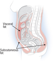
Visceral Fat

Do you see the difference between visceral fat and subcutaneous fat? Visceral fat surrounds the abdominal organs, located behind the abdominal cavity and it is linked to metabolic disturbances, is even associated with breast cancer.

Enough with the bad news, I know.

So what can you do about your chubby belly?

Not that hard to combat abdominal fat gain, it turns out. Regular participation in physical activity of moderate intensity at least 30 minutes per day (or perhaps up to 60 minutes to lose it.) can control your weight. One significant difference is, for example; jogging 12 miles/week can help you prevent visceral fat, whilst jogging 20 miles/week can help you get rid of your visceral fat. Also, weight training twice a week can reduce the propotion of body fat and successfully keep off visceral fat.

Pro tip: Abdominal muscles ≠ visceral fat: spot exercises tighten abdominal muscles, but these exercises won’t affect visceral fat.

And diet… Just like it was mentioned, empahasizing complex carbohydrates, such as fruits, vegetables and whole grains, paying attention to portion sizes can most certainly be beneficial. Don’t drastically cut calories and force your body into starvation mode. If you slow your metabolism, it will paradoxically cause your metabolism to store more fat later on.

One last thing, I’ve been reading a lot of crap about “clean eating” as of late, they suggest that it is a bad thing because “clean eating” can lead to obsessions and eating disorders. True but mostly false.

The thing is – what leads a person to develop obsessions, an eating disorder or any kind of mental disorder are conceptualized as the disorders of brain circuits that are results of a developmental process. Any mental disorder can be a result of nature or nurture, or usually both. (nature: genetics, nurture: life experiences) Life experiences alone aren’t likely to lead a healthy person to develop a mental disorder,  “clean eating” might only trigger or worsen the symptoms of the existing mental disorder. Meal planning will lead my Mom to have  an eating disorder? Come on. Do not generalize anything, it is stupid, every individual is different, do you even know of  my genetic mutations, how can you decide on my genetic mutations’ behalf? Or if you say clean eating is associated with genetic mutations, then prove it. Pffft.

(Genetic mutations: A mental illness is most likely to arise from a genetic mutation. So is Cancer, etc. Be patient, I will eventually write about genetic mutations. I wish I had enough hours in a day to blog everyday.)

Practically, unless you have some metabolic disorder, you can eat whatever you want but be smart enough to ensure a balance of nutritional intake. Balance always is the key to happiness. Keep your life and your meals in balance, exercise and you will be succeeded.

Case Study: Is It Anemia?

An 84-year-old asymptomatic man has been seeing his physician regularly and has been repeatedly found, over several years, to have a hemoglobin level of 12 to 13 g/dl without any symptoms of anemia.

On two previous occasions, iron studies revealed normal serum iron and transferrin saturation but elevated ferritin levels (800 to 900 range). Fecal occult blood was negative and a screening colonoscopy five years ago was normal.
The patient now makes an appointment, earlier than his scheduled routine follow-up, and complains of fatigue and dyspnea with effort, accompanied by a vague sensation of chest pressure. Physical examination is normal except for a symmetrically enlarged prostate without nodules. An initial CBC revealed a hemoglobin of 10 g/dl, hematocrit 30%, white blood cell count 6,400/microliter and platelet count of 140,000/microliter.
Which of the following statements are true and which are false?
A – He appears to have a low-risk progressive anemia (possibly a myelodysplastic syndrome) and should be started on an erythropoietin preparation by injection.
B – A serum erythropoietin level should be determined first and, if it is less than 200 mU/ml, erythropoietin injections should be started.
C – He should be transfused at once to achieve a hemoglobin level of 12 g/dl.
D – He needs an immediate bone marrow aspiration and biopsy.
E – The persistence and progression of anemia, along with a low borderline platelet count, suggest a bone marrow disorder.
F – A bone marrow karyotype of del(5q) alone would be an indicator of a poor prognosis.
G – A reticulocyte count would help determine whether or not the anemia results from a failure of red blood cell production.
H – An elevated direct bilirubin would suggest that there is hemolysis.
I – The absence of splenomegaly is evidence against a primary bone marrow disorder.
J –  Examination of the peripheral blood smear may add useful information about his diagnosis.
(A – False,  B – False, C – False, D – False, E – True, F – False, G – True, H – False, I – False, J – True)
A – False – It is true that the patient has a mild progressive anemia, but it is premature to conclude that it is a myelodysplastic syndrome (MDS) and that it is necessary to start an erythropoietin preparation.  It is premature to treat this patient with an erythropoietin preparation until the etiology of the anemia is established. If an appropriate use for erythropoietin is established, guidelines differ on whether it is necessary to determine a serum erythropoietin level to treat patients with MDS or cancer chemotherapy related anemia.
B – False – However, for treatment of symptomatic anemia secondary to myelodysplastic syndromes, it is suggested measuring the erythropoietin level and using erythropoietin only if the serum level is less than or equal to 500 milliunits/ml.
C – False – An immediate transfusion is not called for. In general, tissue oxygenation, heart rate, blood pressure and the nature of bleeding or hemolysis can provide good clinical information upon which to make a decision to transfuse a patient with red blood cells. We have the following clinical information — a slowly progressive anemia not suggestive of rapid bleeding or hemolysis and the “vague sensation of chest pressure” that has not been determined to be from tissue hypoxia. If the symptoms are caused by myocardial ischemia, red blood cell transfusion up to a hemoglobin level of 12 g/dl might be indicated.
D – False – A bone marrow aspiration is not an immediate need, although it may be an appropriate step if the history and physical examination of the peripheral blood smear, reticulocyte count and stool for occult blood all continue to suggest a myelodysplastic syndrome. It would then be appropriate, together with a bone marrow biopsy, flow cytometry and cytogenetics.  The bone marrow findings would then be used to guide treatment of coexisting causes by replacing iron, folate or B12 if needed.
E – True – When abnormalities of multiple cell lines (RBCs, WBCs and platelets) are found, one must consider an underlying bone marrow disorder.
F- False – While a bone marrow karyotype of del(5q) would indicate the presence of MDS, del(5q) alone is actually a good prognostic indicator, along with normal -Y chromosome alone and del(20q) alone.
G – True – Reticulocytes are red blood cells that still have residual RNA from their maturation in the bone marrow. The RNA shows up in a reticular pattern with special stains giving them their name. They lose this special staining property as they mature over their first day or two in the circulation, so they can be used as a guide to how many red blood cells are being formed. Thus, the reticulocyte count is an index of red blood cell production. The reticulocyte count is often expressed as a percentage of red blood cells, but can also be expressed as an absolute count or a “corrected” count when the percentage is adjusted for anemia.
H- False – Although the bilirubin level may be elevated by hemolytic anemia, it is the indirect bilirubin that is elevated, not the direct bilirubin.
I – False – Splenomegaly may occur with or without a bone marrow disorder and a bone marrow disorder may occur with or without splenomegaly.
J – True – Examination of the blood smear may reveal abnormalities of red blood cell morphology and white blood cell morphology not obvious on a routine CBC.
Next, our elderly patient is referred to a cardiologist, undergoes cardiac catheterization, and is found to have triple vessel plus left main stem coronary artery disease. Urgent coronary artery bypass graft (CABG) surgery is performed.
Indicate which of the following statements or questions are true and which are false?
A –  Collecting a bone marrow specimen at the time of surgery may be useful.
B – The patient should be transfused to maintain a hemoglobin level of 12 g/dl in the immediate post-operative period.
C – He should be started on erythropoietin injections immediately post surgery in anticipation of an inability to recover from surgical blood loss.
D – He should be started on oral iron, folate and vitamin B12 after surgery.
E – The surgery might not have been done if a hematologic diagnosis had been established.
F – His pre-operative history, physical and laboratory evaluation may provide clues to his diagnosis.
G – Once his operative blood loss is replaced with transfusion, his hemoglobin level should remain stable.
H – If an examination of the bone marrow is consistent with MDS, consideration should be given to treatment with an erythropoietin preparation at a dose of 150-300 units/kg/day by subcutaneous injection.
I – If his hemoglobin rises to 15 g/dl after receiving erythropoietin alfa for two to three months, the dose should be decreased, as tolerated, to maintain a hemoglobin level of 12 g/dl.
(A – True , B – False , C – False, D – False, E – True, F – True, G – False, H – True , I – True)
A – True – Collecting a bone marrow specimen is easy and convenient at the time of sternotomy for cardiac surgery. However, the trauma of the sternotomy and poor specimen handling often result in biopsy specimens that are technically poor and difficult to interpret. A properly collected biopsy, with samples for flow cytometry and cytogenetics, is often much more rewarding.
B – False – Once the heart has been revascularized, the threat of critical myocardial ischemia secondary to anemia is removed and the patient usually will easily tolerate moderate anemia.
C – False – Although his bone marrow might not be able to respond adequately to the blood loss of surgery, it is not certain and any decision to start an erythropoietin preparation should be deferred until there is clear evidence that it is needed.
D – False – In view of his elevated ferritin, giving the patient oral iron that he may not be able to utilize in the formation of new red blood cells would be an error that could increase his risk of developing hemosiderosis as a later complication of a transfusion dependent MDS. Vitamin B12 or folate supplementation would have less potential for causing harm but should probably be given only if there is a clear indication for their need.
E –  True – If an MDS had been identified and his anemia successfully treated, he might not have developed cardiac symptoms.
F – True – A full work-up might have led to a definitive hematologic diagnosis. It would have been helpful if this were available before a decision was made about heart surgery.
G – False – It should be anticipated that the anemia will recur and he should be monitored so that intervention can be carried out as needed.
H/I – True – The recommendations for epoetin alfa are correct.

How the ultimate smart phone sends text messages: Neurotransmitters.

I truly believe that the idea of Internet was inspired by the Central Nervous System. Though, I may be biased because I pretty much believe that everything is inspired by Central Nervous System!

Let’s get the party started? Alright.

What is a neurotransmitter? A chemical in your brain which transmits signals from a neuron to a target across a synapse.

Synapse? Look here:

Synapse

Chemical synapses allow neurons to form circuits and CNS to connect and control the body. Your brain is estimated to contain 100-400 trillion synapses. Electrical and immunological synapses, such as Cytokine network exist but today I only talk about the chemical synapses.

Most synapses connect axons to dendrites, you can see the image above. However, there are other types of connections, such as axon-to-axon, axon-to-soma, dendrite-to-dendrite.

Soma, dendrite and axon? Soma is a body cell. Dendrite can be described as a signal receiver and axon can be the projection which conduct the signal. A neuron cell has protrusions. The small protrusions are called “dendrites”  and on their ends the are the “synapses” that receive chemical signals from other neuron cells. The long protrusions are called the “axons”, they are nerve endings and can release chemical signals to be received on the synapses of neurons in its neighborhood.

Before we start, we really need to know what a NEURON really is;

  1. Glial cells are responsible for neurons. (Probably,  you’ve heard me say “Glioma” for about million times. Now you also know Glioma arises from glial cells.)
  2. Neurons make up the gray matter of the nervous system.
  3. Neurons are excitable cells. Excitable cells can be stimulated to create a electric current.
  4. Neuron processes running through the CNS form tracts of white matter; outside the CNS they form the peripheral nerves.
  5. The neuron cell body contains a large round nucleus, this nucleus is surrounded by cytoplasm, cytoplasm is riddled with Nissl bodies, Nissl bodies are like endoplasmic reticulum and everyone knows what an endoplasmic reticulum is. This is the one and only thing you always remember from your biology classes in high school.
  6. Neuron

    As you can see, neurons have many dendrites but only one axon. Axons carry impulses away from the nerve cell body and dendrites conduct electrical currents toward the cell body. Axon terminals are enlarged axon endings used to make contacts with other nerve cells.

Let me explain the process in general:

  • Neurons receive signals in two forms; a) chemical changes, which happens via neurotransmitters, b) physical changes, such as touch receptors, photoreceptors. (Photoreceptor: A photosensitive cell that is responsible for detecting light and, therefore, enables us to see.)
  • Signals are sent from one neuron to another by jumping across a synapse.
  • Presynaptic neuron, the neuron sending the signal has small, membraine-bound compartments that package neurotransmitters within the cell, and voltage-gated protein channels that open or close in response to charge difference at the membrain, specifically, presynaptic neuron has calcium channels that allow calcium into the cell.
  • Postsynaptic neuron, the neuron receiving the signal has receptor molecule, a protein that recognizes a specific 3-dimensional shape.
  • When an action potential begins in a neuron, it travels down the axon. Actually, it all begins with a kiss called action potential, the changes which occur during nerve impulse propagation. Neurons are coated with myelin, a sheath, a collection of lipid fats and proteins that is produced by oligodendrocytes, these sheats act as insulators, preventing ion leakage and hence increasing speed of nerve impulse propagation helping electrical current flow down the axon, but the axons have “Nodes of Ranvier” in their “myelinated coats” every 0.2-2 mm, the places where ion transfers take place, action potentials traveling down the axon “jump” from node to node, it’s called saltatory conduction which means “to leap.” (Further knowledge; Multiple Sclerosis (MS) results in the gradual destruction of these myelin sheaths.)
  • When the action potential reaches the axon terminal, calcium channels open and calcium ions rush into the neuron.
  • The neuron MAKES and STORES neurotransmitters (Neurotransmitters are finally mentioned, I know.) in vesicles.
  • When calcium binds to the vesicles, the vesicles carry neurotransmitter toward the presynaptic (neuron’s) membrane.
  • When the vesicles contact the axon terminal membrane, the neurotransmitter is released into the synaptic cleft. (Synaptic cleft: A microscopic gap that exists between the neurons.)
  • Neurotransmitter diffuses across the synaptic cleft and bind to receptors on the postsynaptic neuron.
  • The postsynaptic neuron receptors are activated, in this case, these receptors allow Na in the neuron faciliated diffusicon, causing an action potential to start in the post synaptic membrane.
  • Neurotransmitters are released from receptors and diffuse back into the synaptic cleft.
  • Vesicles recycle some neurotransmitters to prepare the neuron for its next action potential.

And this is pretty much it. You just made a connection because you learned somethin new, maybe even changed a synapse. And you had fun?

Tip: Some drugs, such as caffeine, are stimulants that decrease the postsynaptic neuron’s threshold and make it more irritable. Others block transmission by interfering with the release of neurotransmitter or by the synaptic knob. Some of these drugs are used as painkillers, etc.

Now that we know neurotransmitters allow the transmission of signals from one neuron to the next across synapses, are also found at the axon endings of motor neurons, produced by pituitary and adrenal glands, etc, it’s almost time for us to finally learn what certain neurotransmitters DO.

There are many types of chemicals that act as neurotransmitter substances, let’s make our list and I will review some of the most important neurotransmitters.

Read more of this post